B-cell intestinal lymphoma with Mott cell differentiation in a 1-year-old miniature Dachshund.
Słowa kluczowe
Abstrakcyjny
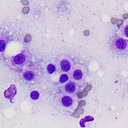
A 1-year-old intact female miniature Dachshund was presented with hematochezia, vomiting, and diarrhea of more than 1-week duration. An abdominal mass was palpated, which at exploratory surgery was found to be a 7-cm-long thickened section of ileum. The thickened ileum was resected. Impression smears revealed numerous small- to medium-sized lymphocytes, with a smaller number of cells resembling Mott cells. The Mott-like cells contained multiple pale vacuoles that were positive for periodic acid-Schiff (PAS) in wet-fixed smears, consistent with Russell bodies. Histologic evaluation of the surgically excised ileum revealed 2 populations of neoplastic lymphoid cells. The majority were uniform medium-sized lymphocytes with hyperchromatic oval or round nuclei and inconspicuous nucleoli. The remaining cells resembled Mott cells, which contained several PAS-positive eosinophilic globules in the cytoplasm, occasionally compressing the nucleus. The majority of neoplastic cells stained positively for vimentin, CD20, CD79a, and Pax-5, but were negative for CD3 and lysozyme; 43.5% of cells stained positively for Ki-67. The Mott cells were strongly positive for immunoglobulin but were negative for Pax-5. Using electron microscopy, a homogenous substance of intermediate electron density was observed frequently in the cisternae of rough endoplasmic reticulum in the cytoplasm of the Mott cells, and rarely in the perinuclear cisternae of the lymphoid cells, corresponding to the site of immunoglobulin staining. Monoclonal rearrangement of immunoglobulin heavy-chain (IgH) gene was observed by PCR testing for lymphocyte-antigen receptor rearrangement. The morphologic features, immunophenotype, and IgH gene rearrangement verified the lymphoid cells were neoplastic (mature cell type) and had a B-cell phenotype, with evidence of immunoglobulin production and differentiation into Mott cells. This case was unusual because of the age of the dog and because most intestinal lymphomas are T-cell phenotype. The Mott cell morphology also differed from typical mature B-cell lymphoma types and may be a unique B-cell lymphoma variant.